Wednesday, October 23, 2013

Plant Cell

Plant Cell Anatomy
Animal Cell PrintoutBacterium Cell Printout

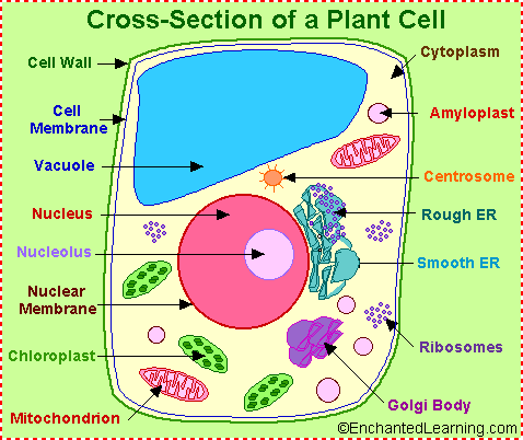
The cell is the basic unit of life. Plant cells (unlike animal cells) are surrounded by a thick, rigid cell wall.
Plant cell anatomy

The following is a glossary of plant cell anatomy terms.
amyloplast - an organelle in some plant cells that stores starch. Amyloplasts are found in starchy plants like tubers and fruits.
ATP - ATP is short for adenosine triphosphate; it is a high-energy molecule used for energy storage by organisms. In plant cells, ATP is produced in the cristae of mitochondria and chloroplasts.
cell membrane - the thin layer of protein and fat that surrounds the cell, but is inside the cell wall. The cell membrane is semipermeable, allowing some substances to pass into the cell and blocking others.
cell wall - a thick, rigid membrane that surrounds a plant cell. This layer of cellulose fiber gives the cell most of its support and structure. The cell wall also bonds with other cell walls to form the structure of the plant.
centrosome - (also called the "microtubule organizing center") a small body located near the nucleus - it has a dense center and radiating tubules. The centrosomes is where microtubules are made. During cell division (mitosis), the centrosome divides and the two parts move to opposite sides of the dividing cell. Unlike the centrosomes in animal cells, plant cell centrosomes do not have centrioles.
chlorophyll - chlorophyll is a molecule that can use light energy from sunlight to turn water and carbon dioxide gas into sugar and oxygen (this process is called photosynthesis). Chlorophyll is magnesium based and is usually green.
chloroplast - an elongated or disc-shaped organelle containing chlorophyll. Photosynthesis (in which energy from sunlight is converted into chemical energy - food) takes place in the chloroplasts.
christae - (singular crista) the multiply-folded inner membrane of a cell's mitochondrion that are finger-like projections. The walls of the cristae are the site of the cell's energy production (it is where ATP is generated).
cytoplasm - the jellylike material outside the cell nucleus in which the organelles are located.
Golgi body - (also called the golgi apparatus or golgi complex) a flattened, layered, sac-like organelle that looks like a stack of pancakes and is located near the nucleus. The golgi body packages proteins and carbohydrates into membrane-bound vesicles for "export" from the cell.
granum - (plural grana) A stack of thylakoid disks within the chloroplast is called a granum.
mitochondrion - spherical to rod-shaped organelles with a double membrane. The inner membrane is infolded many times, forming a series of projections (called cristae). The mitochondrion converts the energy stored in glucose into ATP (adenosine triphosphate) for the cell.
nuclear membrane - the membrane that surrounds the nucleus.
nucleolus - an organelle within the nucleus - it is where ribosomal RNA is produced.
nucleus - spherical body containing many organelles, including the nucleolus. The nucleus controls many of the functions of the cell (by controlling protein synthesis) and contains DNA (in chromosomes). The nucleus is surrounded by the nuclear membrane
photosynthesis - a process in which plants convert sunlight, water, and carbon dioxide into food energy (sugars and starches), oxygen and water. Chlorophyll or closely-related pigments (substances that color the plant) are essential to the photosynthetic process.
ribosome - small organelles composed of RNA-rich cytoplasmic granules that are sites of protein synthesis.
rough endoplasmic reticulum - (rough ER) a vast system of interconnected, membranous, infolded and convoluted sacks that are located in the cell's cytoplasm (the ER is continuous with the outer nuclear membrane). Rough ER is covered with ribosomes that give it a rough appearance. Rough ER transport materials through the cell and produces proteins in sacks called cisternae (which are sent to the Golgi body, or inserted into the cell membrane).
smooth endoplasmic reticulum - (smooth ER) a vast system of interconnected, membranous, infolded and convoluted tubes that are located in the cell's cytoplasm (the ER is continuous with the outer nuclear membrane). The space within the ER is called the ER lumen. Smooth ER transport materials through the cell. It contains enzymes and produces and digests lipids (fats) and membrane proteins; smooth ER buds off from rough ER, moving the newly-made proteins and lipids to the Golgi body and membranes
stroma - part of the chloroplasts in plant cells, located within the inner membrane of chloroplasts, between the grana.
thylakoid disk - thylakoid disks are disk-shaped membrane structures in chloroplasts that contain chlorophyll. Chloroplasts are made up of stacks of thylakoid disks; a stack of thylakoid disks is called a granum. Photosynthesis (the production of ATP molecules from sunlight) takes place on thylakoid disks.
vacuole - a large, membrane-bound space within a plant cell that is filled with fluid. Most plant cells have a single vacuole that takes up much of the cell. It helps maintain the shape of the cell.

Sunday, October 20, 2013

The Structure of Uman Blood

Red blood cells have a unique shape and inner components that allow
them to efficiently transport oxygen and direct the elimination of carbon dioxide.

Bone Marrow is the "blood cell factory" which is found filling up the cavities of bones. All blood cells originate and are produced from a single "stem cell" whose progeny grow and mature into different types of blood cells. This stem cell can and does renew itself as required by our body.

Red blood cells (erythrocytes) carry oxygen from your lungs to all parts of your body. If you don't have enough red blood cells you have anemia.

White blood cells (leucocytes) are the body's infection fighters. There are three main groups of white blood cells: granulocytes, lymphocytes and monocytes. Their job is to rid your body of disease-causing bacteria, viruses, fungi and to destroy the body's dead or defective cells. If we do not have enough white blood cells we are at risk of catching all types of infections.

Platelets are small cells that prevent bleeding and makes blood clot following an injury. When a blood vessel is damaged or cut, platelets rush to the area and clump together to plug the bleeding site. If we do not have enough platelets easy bruising, nose bleeds, prolonged bleeding from cuts, or internal bleeding from the bowel or bladder may occur.

The trillions of cells that compose the human body rely on red blood cells, a.k.a. erythrocytes to supply them with oxygen and help direct the elimination of waste carbon dioxide gas. Red blood cells have a unique structure and properties that allow them to fulfill these crucial missions. Red blood cells (RBCs) are the primary cells in human blood. They are biconcave discs, having a depressed center on both sides. These depressed centers allow the cells to have more cell membrane surface which can be exposed to diffusing oxygen while transiting the lungs. This structure also allows them to be more flexible when negotiating tight passages.

RBCs are about 7.8 micrometers in diameter (A micrometer is 1/1,000,000 of a meter). They have a flexible nature that allows them to bend and bounce back their original shape. This comes in handy when they must squeeze through the minute capillary alleyways between cells in the tissues.

Hemoglobin

Unlike most cells, mature RBCs of humans don’t have nuclei, mitochondria, or other organelles. Instead, they are packed full of a special substance called hemoglobin (Hgb), a complex molecule composed of protein and iron. Hgb is responsible for picking up oxygen, which diffuses across membranes in the small vessels of the minute lung sacks called alveoli. Hgb holds onto oxygen until it reaches areas of the body where it is in low concentration and then releases it to diffuse into local tissues.

RBCs are passive in nature, being swept along by the blood. They sustain their meager energy needs by a form of anaerobic respiration. Since they don’t have mitochondria, there are no worries they might gobble up the oxygen they are charged with transporting.

Management of Carbon Dioxide

RBCs also contain an enzyme called carbonic anhydrase which takes carbon dioxide and water and catalyzes the creation of a stable molecule called bicarbonate (HCO3-). Bicarbonate dissolves much better than carbon dioxide in the fluid part of blood and great quantities can be transported this way without needing to be in the small red blood cells.

Structural Abnormalities of RBCs

Sometimes structural changes occur in RBCs that indicate a possible pathological condition. For instance, one may observe enlarged RBCs with folate or vitamin B12 deficiency (macrocytic anemia). Small RBCs may be observed with iron deficiency or chronic blood loss (microcytic anemia). Sickle cell anemia is a condition where a genetic flaw leads to defective hemoglobin that changes shape under certain conditions. This shape change induces the cell to lose its original biconcave disc confirmation and take on a more sickled appearance.


Classification & Structure of Blood Vessels

Blood vessels are the channels or conduits through which blood is distributed to body tissues. The vessels make up two closed systems of tubes that begin and end at the heart. One system, the pulmonary vessels, transports blood from the right ventricle to the lungs and back to the left atrium. The other system, the systemic vessels, carries blood from the left ventricle to the tissues in all parts of the body and then returns the blood to the right atrium. Based on their structure and function, blood vessels are classified as either arteries, capillaries, or veins.

Arteries

Arteries carry blood away from the heart. Pulmonary arteries transport blood that has a low oxygen content from the right ventricle to the lungs. Systemic arteries transport oxygenated blood from the left ventricle to the body tissues. Blood is pumped from the ventricles into large elastic arteries that branch repeatedly into smaller and smaller arteries until the branching results in microscopic arteries called arterioles. The arterioles play a key role in regulating blood flow into the tissue capillaries. About 10 percent of the total blood volume is in the systemic arterial system at any given time.

Illustration on an artery wall

The wall of an artery consists of three layers. The innermost layer, the tunica intima (also called tunica interna), is simple squamous epithelium surrounded by a connective tissue basement membrane with elastic fibers. The middle layer, the tunica media, is primarily smooth muscle and is usually the thickest layer. It not only provides support for the vessel but also changes vessel diameter to regulate blood flow and blood pressure. The outermost layer, which attaches the vessel to the surrounding tissue, is the tunica externa or tunica adventitia. This layer is connective tissue with varying amounts of elastic and collagenous fibers. The connective tissue in this layer is quite dense where it is adjacent to the tunic media, but it changes to loose connective tissue near the periphery of the vessel.

Veins

Veins carry blood toward the heart. After blood passes through the capillaries, it enters the smallest veins, called venules. From the venules, it flows into progressively larger and larger veins until it reaches the heart. In the pulmonary circuit, the pulmonary veins transport blood from the lungs to the left atrium of the heart. This blood has a high oxygen content because it has just been oxygenated in the lungs. Systemic veins transport blood from the body tissue to the right atrium of the heart. This blood has a reduced oxygen content because the oxygen has been used for metabolic activities in the tissue cells.

Illustration of the walls of a vein

The walls of veins have the same three layers as the arteries. Although all the layers are present, there is less smooth muscle and connective tissue. This makes the walls of veins thinner than those of arteries, which is related to the fact that blood in the veins has less pressure than in the arteries. Because the walls of the veins are thinner and less rigid than arteries, veins can hold more blood. Almost 70 percent of the total blood volume is in the veins at any given time. Medium and large veins have venous valves, similar to the semilunar valves associated with the heart, that help keep the blood flowing toward the heart. Venous valves are especially important in the arms and legs, where they prevent the backflow of blood in response to the pull of gravity.

Copy Right ILMU-online.blogspot.com